Close

Cerebellar Pathways

*

*

Diagram of the cerebellar nuclei showing the fastigial, interposed (globose and emboliform) and dentate nuclei.

Functions of the Cerebellar lobes and nuclei

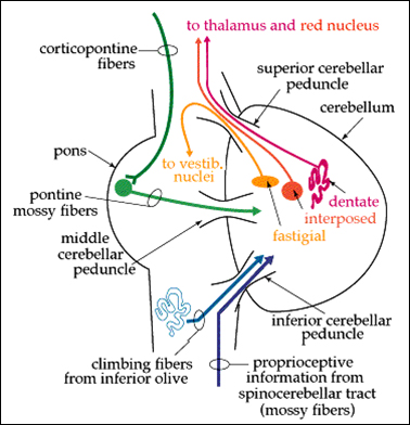
The diagram (above, left) shows the main connections of the cerebellum.

The inferior cerebellar peduncle carries axons from the spinal cord (the spinocerebellar and cuneo-cerebellar tracts; and in addition a major pathway form the contralateral olivary nuclei. The axons of these fibres are known as 'climbing fibres', and their functions are discussed in the section of Cerebellar Physiology.

The middle cerebellar peduncle consists largely of the axons of pontine nuclei that carry a copy of messages sent down the corticospinal pathway; this occurs because the cortico-bulbar (cortico-pontine) neurones synapse on pontine nuclei. The terminals of these axons have a characteristic structure and are know as the 'mossy fibres'.

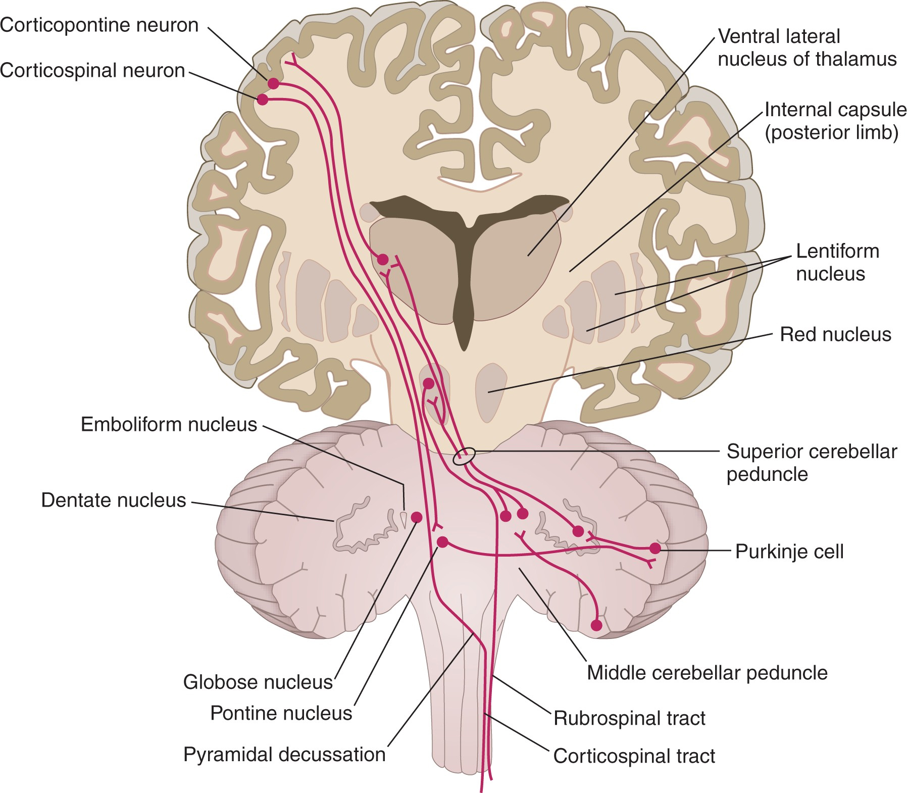
The superior cerebellar peduncle is the main efferent pathway from the cerebellum, and carries the axons of the deep cerebellar nuclei (shown in the diagram on the right). The fastigial nuclei belong to the oldest part of the cerebellum and project to the vestibular nuclei. The dentate and interposed nuclei project rostrally to synapse in the red nucleus of the midbrain and the thalamus (see the diagram below, right).

The vermis connects with pathways passing from the brainstem to the spinal cord (bulbo-spinal pathways) that descend to motoneurones and influence muscle tone. Two groups of bulbo-spinal pathways - medial and lateral are influenced by the vermis. This region also receives proprioceptive information from the spinal cord.

The hemispheres of the cerebellum project to the motor areas of the cerebral cortex, and are concerned with motor planning and execution. These connections were developed along with the growth of the cerebral cortex and cerebellum during the evolution of the upright posture.

The diagram opposite shows the connections of the neocerebellum and the neocortical regions concerned with motor control .

Let's start with the efferent pathways. The Dentate nucleus is the largest of the deep cerebellar nuclei and deals with information processed by the neocerebellum and sends its infomation to the red nucleus, thalamus and neocortex. These pathways are vary much involved with the maintenance of the upright posture, and the coordination of movements.

The neurones of the red nucleus receive inputs from the neocerebellum and pass it on the (a) the thalamus, (b) the spinal cord, and (c) the inferior olivary nucleus. The rubro-spinal tract accompanies the corticospinal tract within the cord.

The neocerebellum receives its major inputs from the neocortex via the pontine nuclei.

Thus the functions of the neocerebellum are concerned with receiving a copy of the motor commands via the pontine nuclei, and information from proprioceptors and vestibular apparatus. This allows the organ to function as a comparator producing an error signal that is relayed back to the cortex.

Sometimes the efferent pathways from the cerebellum pathways are referred to as the cerebello-rubro-thalamo-cortical pathway - which originates in the cerebellum and reaches the cortex after synapsing in the red nucleus in the tegmentum of the midbrain and then also in the thalamus.

The cortico-pontine cerebellar pathway refers to the chain of neurones consisting of the cerebro-pontine fibres followed by the ponto-cerebellar fibres.


The diagram shows the corticospinal neurones sending axons to the spinal cord.

  1. The cortico-pontine fibres send a similar message to pontine neurones that project to the cerebellar cortex.
  2. The Purkinje cells process that information and the result is sent to the Dentate nucleus (the main efferent pathways from the cerebellum).
  3. The dentate neurones send their mesages to the thalamus, and some relay in the red nucleus.
  4. Finally the cortex receives the message from the thalamus.
Note that the red nucleus also sends information to the spinal cord using the rubro-spinal tract, and also the contralateral inferior olive (see below).

Rubro-Olivary Pathways

The Inferior Olive is another source of axons that project to the cerebellum.

It is known to have inputs from the red nucleus (see the diagram opposite) and probably also from the motor cortex, via the corticospinal tract.

Note that the red nucleus is one of the sites that receives an input from the cerebellum, and the pathway from the red nucleus to the olive may allow the cerebellum to receive some feedback about its own output, although this function is not fully understood.

So although the rubro-olivary tract is outside the cerebellum it could potentially play a part in its function.


*
*

Pathways through the flocculonodular lobe are associated with the Vestibular apparatus and nuclei. The Fastigial Nucleus has two parts concerned with vestibular function (a) one is concerned with the vestibulo-spinal system, and (b) the other has to do with vestibulo-ocular reflexes.

*

The Spino-Cerebellar pathways pass through the spinocerebellum and project through the interposed nuclei to the red nucleus and the motor areas of the cortex. There are several maps of the body surface on the spino-cerebellar cortex, and the red nucleus (and others related nuclei) project to the spinal cord and the inferior olivary nucleus.

The inferior olivary nucleus is of significance in cerebellar function because it is the origin of the powerful climbing fibres that are central to each cerebellar module or microzone.

The Inferior Olive

This transverse section of the medulla shows the close relationship between the pyramids (cerebrospinal fasciculus) and the inferior olive, which causes and olive-shaped bulge on the surface of the medulla.

The cell bodies are arranged in a 'U' shape with its centre directed towards the contralateral inferior cerebellar peduncle (restiform body).

Note the position of the olive and the olivocerebellar fribres projecting from its centre to the opposite side of the cerebellum

*

There are some colorful names associated with the brainstem motor pathways.

  • The Substantia Nigra is black, due to the presence of melanin.
  • The Red Nucleus is pink, due to the large capillary network in that area, possibly indicating the importance of bood flow to this active region.
  • The Locus Coeruleus, a noradrenergic nucleus in the upper pons, is so named because it is bluish in colour; apparently this is associated with light scattering by neuromelanin. This nucleus sends its axons throughout the cerebellum.
imueos.blogspot.co.uk
www.dizziness-and-balance.com
www.lookfordiagnosis.com
what-when-how.com
Gray's Anatomy

Top

Close

© HumanPhysiology.Academy 2014-2015